Image Analysis Core
Core Leader: Ron Korstanje
Phone: 207-288-6992
email: ron.korstanje@jax.org
The overall goal of the Image Analysis Core is to develop and provide resources for the geroscience community to aid in computer-assisted histopathological analysis and discovery of age-related histological features. Recently, the NIA-funded Geropathology Research Network (GRN), established to enhance the translational value of geropathology for preclinical research studies in anti-aging clinical trials, developed and validated a grading system, designated the geropathology grading platform (GGP), for quantification and comparison of histological lesion scores in tissues from aging mice. While implementation of this grading platform by a trained pathologist may be feasible for experiments with small numbers of animals, an automated approach is necessary for experiments consisting of large sample numbers. An automated approach that can provide unbiased analysis of large sample numbers will lead to a more timesaving and cost-effective analysis and generation of more robust data. A quantitative image analysis pipeline that uses machine learning to accurately identify specific features in scanned slides of stained kidneys was recently developed. This quantitative tool can be easily adjusted to allow quantification using the GGP.
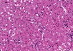
Our pipeline can easily distinguish between young kidney tissue
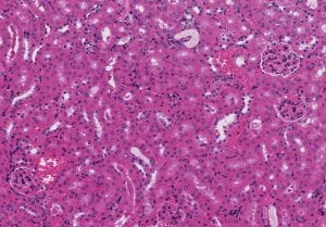
and old kidney tissue.
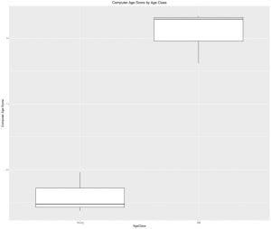
Not only can we make these distinctions, we can quantify and measure old and young tissues.
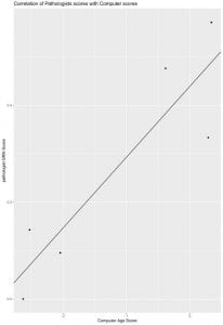
This computerized aging score has a high correlation with tissues pathologists score using the GRN platform.
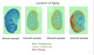
Looking to further refine and understand where aging happens, we can “paint” our age scores on tissues and look at where aging is happening.
Tutorials about how to apply these tools to your own slides and how to visualize and look at the differences can be found at geropathology-imaging.org.